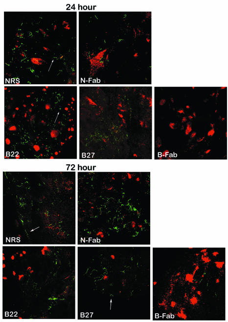
FIG. 4.
OspB F(ab)2 fragments interfered with the attachment and colonization of B. burgdorferi within I. scapularis. The distribution of B. burgdorferi within the I. scapularis gut 24 and 72 h after feeding is shown. Nymphal ticks fed on B. burgdorferi-infected mice that had been treated with either normal rabbit serum (NRS), F(ab)2 fragments from normal rabbit serum (N-Fab), or OspB MAb B22 or B27 or F(ab)2 fragments prepared from polyclonal anti-OspB sera (B-Fab). The spirochetes (arrows) were stained with a FITC-labeled goat anti-B. burgdorferi antibody (green), and the nuclei of the gut epithelial cells were stained with propidium iodide (red). Images were recorded at ×400 magnification and are presented as a merged image for clarity.

