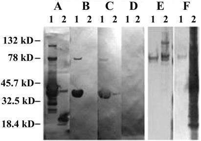
FIG. 3.
Western blotting of P. carinii antigens. (A to D) Western blots of A121-82 (lanes 1) and the thioredoxin fusion partner alone (lanes 2) with (A) anti-V5 epitope tag MAb, (B) 4F11(G1), (C) anti-P. carinii hyperimmune mouse serum, 1:250 dilution, and (D) pooled normal mouse sera, 1:250 dilution. (E and F) Western blots of MAb 4F11 affinity-purified P. carinii antigen (lane 1) and P. carinii-infected mouse lung homogenate (lane 2) with (E) MAb 4F11 and (F) anti-P. carinii hyperimmune serum, 1:250 dilution. Pooled normal mouse sera did not react with either affinity-purified antigens or total P. carinii-infected mouse lung homogenates (data not shown).

