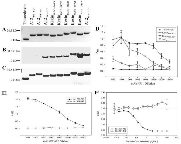
FIG. 5.
Analysis of purified recombinant epitope-thioredoxin fusion proteins. (A) Gel stained with Coomassie blue. (B) Western blot with MAb 4F11(G1), (C) Western blot with anti-V5 epitope tag MAb. (D) ELISA of epitope-thioredoxin fusion constructs with MAb 4F11(G1). Results are plotted as the mean ± standard error of triplicate experiments. (E) ELISA of synthetic P. carinii peptides with MAb 4F11(G1). Results are plotted as the mean ± standard deviation of triplicate experiments. (F) Competitive ELISA with three fold dilutions of synthetic P. carinii peptides as soluble competitors for 4F11(G1) (diluted 1:3,200) binding against plate-bound sonicated mouse P. carinii. Results are plotted as the mean ± standard deviation of triplicate experiments. The dashed line indicates the mean absorbance at 655 nm with no inhibitor and a 4F11(G1) dilution of 1:3,200.

