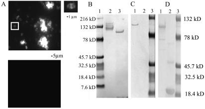
FIG. 6.
MAb 4F11 recognizes surface antigen PspA of S. pneumoniae strain URSP2. (A) (Upper panel) S. pneumoniae was probed with MAb 4F11 and FITC-conjugated secondary antibody; right, enlargement of boxed area showing MAb 4F11 staining of S. pneumoniae diplococcus. (Lower panel) S. pneumoniae probed with isotype-matched MAb 2B5 and FITC-conjugated secondary antibody. (B) Western blot of P. carinii-infected mouse lung homogenates and S. pneumoniae culture lysates probed with MAb 4F11. Lanes: 1, molecular size markers; 2, P. carinii-infected mouse lung homogenate; 3, S. pneumoniae URSP2 lysate. (C and D) Western blots probed with (C) MAb 4F11(G1) and (D) anti-V5 epitope-tagged MAb. Lanes: 1, purified recombinant URSP2 PspA-thioredoxin fusion protein; 2, thioredoxin only; 3, molecular size markers.

