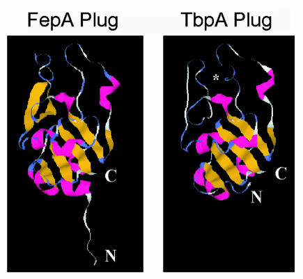
FIG. 8.
Comparison of the three-dimensional structure of the plug region of FepA with the predicted structure of the TbpA plug. The amino termini of the proteins are indicated by N. The carboxy termini are represented by C. The asterisk in the TbpA plug panel indicates the approximate position of Ala110 after the HA epitope was fused in the PHA mutant. β-Strand structure is indicated in yellow, α-helical structure is indicated in red, β-turn structure is indicated in blue, and unstructured sequence is shown in white. A sequence consisting of amino acids 1 to 162 of the processed form of gonococcal TbpA was aligned with the analogous sequence of FepA using the 3D-JIGSAW comparative modeling server.

