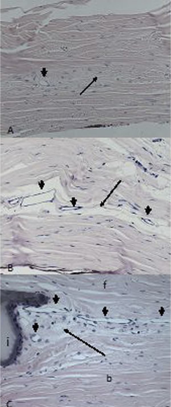
Figure 2.

Light microscopy analysis (H&E ×200). (A) Scar tissue (thin arrow) with single vessel (thick arrow) in operated sclera of eye without implant. (B) Adipose tissue (thin arrow) with vessels (thick arrows) filling space left after absorption of implant. (C) Posterior pole of non-absorbable implant with scar tissue (thin arrow), single macrophages, and vessels (thick arrows) within triangular space limited by implant (i), scleral flap (f) and bed (b).
