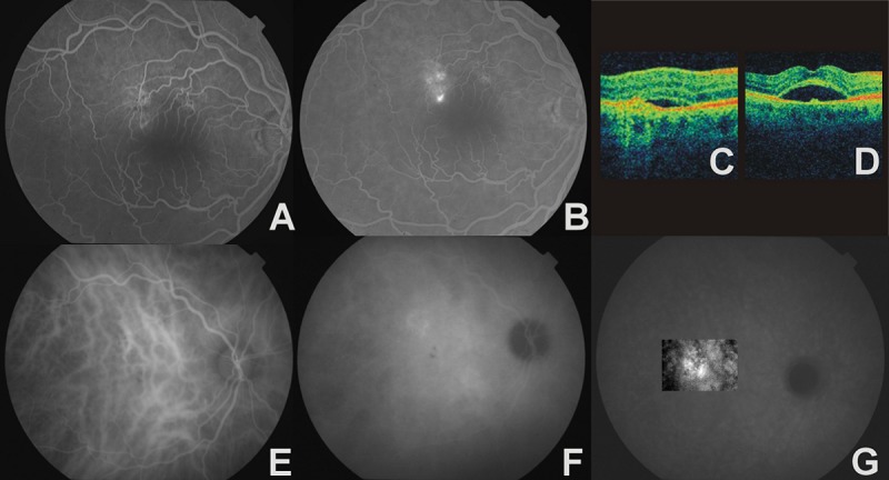
Figure 2.

The right eye of a 65-year-old man with chronic CSC and CNV. (A) – early phase AF, window defect of RPE, (B) – late phase AF, focal leakage points, (C) –4OCT, thickening of RPE/choriocapillaris complex with serous retinal detachment, (D) – OCT, neurosensory retina detachment with bulge protruding from the RPE, (E) – early phase ICGA, venous dilatation, (F) – midphase ICGA, zones of choroidal hyperpermeability, (G) – late phase ICGA, ill-defined, late-staining area that confirms occult CNV.
