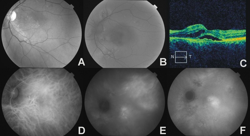
Figure 3.

The left eye of a 54-year-old female with bilateral chronic CSC and CNV. (A) – red-free photograph, hard exudates in macula region, (B) – fundus autofluorescence image, multifocal areas of hyper- and hypoautofluorescence showing RPE abnormality, (C) – OCT, irregular thickening of RPE/choriocapillaris complex with serous retinal detachment, intraretinal edema, (D) – early phase ICGA, areas of hypoperfusion and venous vessels dilatation, (E) – midphase ICGA, multiple zones of choroidal hyperpermeability, (F) – late phase ICGA, ill-defined area of late hyperfluorescence in place of occult CNV.
