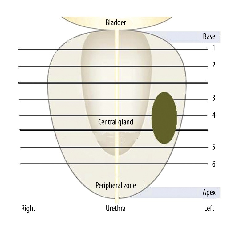
Figure 4.
Ultrasound elastography data collection process using the sextant approach; RF data was acquired in axial planes (1–6) from the gland’s base towards the apex. For illustration purposes, a lesion is outlined in the left mid section, peripheral zone of the specimen, similar with the case of specimen #3.

