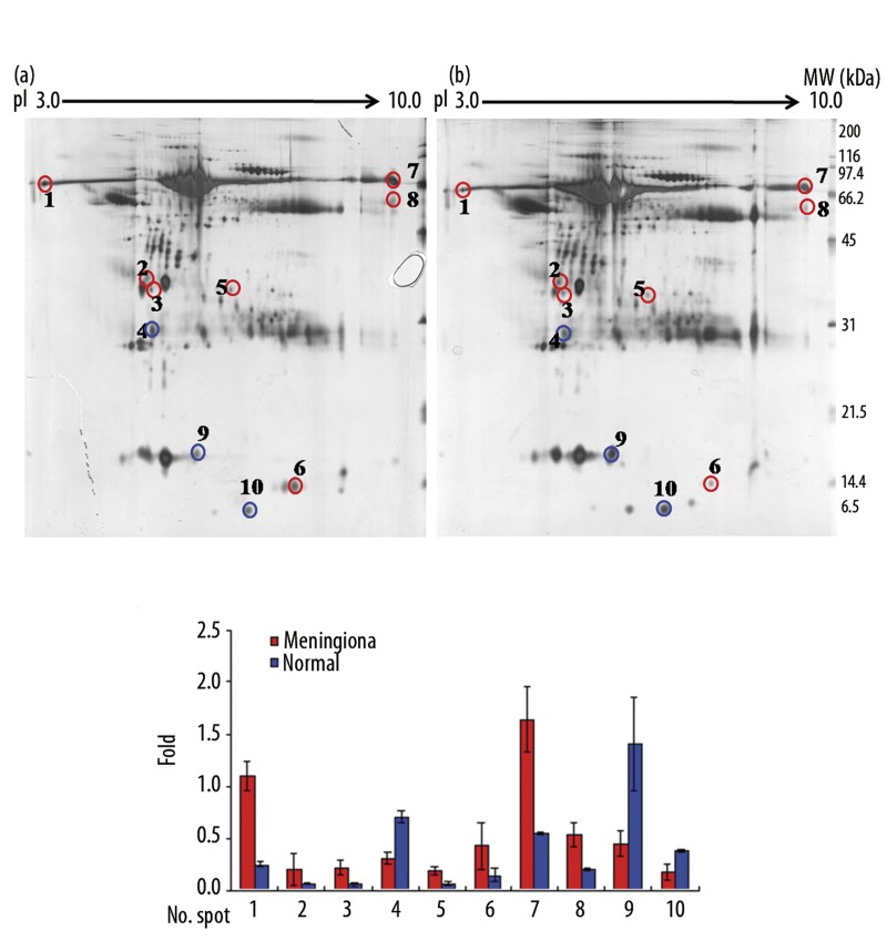
Figure 1.
2-D gel electrophoretic separation of a human CSF proteome. Approximately 150 μg of protein was used. (A) Acetone-precipitated CSF samples from patients with (a) meningioma, and (b) from patients with a non-brain tumor were analyzed by 2-DE analysis using IEF strips, pH 3–10, and 12.5% SDS-PAGE. Spots were visualized by silver staining and spot patterns were analyzed using Progenesis workstation version 2005 software. Samples were run in triplicate. (B) Representative quantifications of CSF proteins in meningioma and non-brain tumor patients. The gels shown represent one of triplicate samples.

