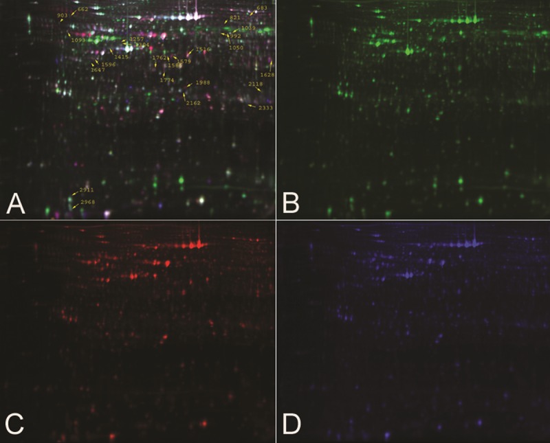
Figure 1.

Representative DIGE fluorescence images of T24 cells with and without siRNA. There are twenty-five differential expressed proterins spots we found, as labeled in the image. (A) Overlays of the Cydye-labeled images; (B) Cydye-labeled image of sample of CON group; (C) Cydye-labeled image of sample of siRNA group; (D) Cydye-labeled image of internal standard.
