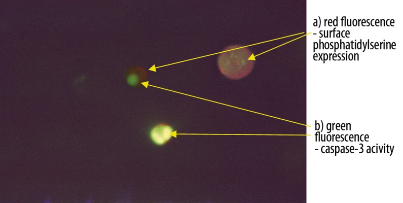
Figure 3.

Dual staining of PMNs with NucView488 Caspase-3 and sulforhodamine101-annexinV after 18 hours of culture. The image shows 3 features of apoptosis: phosphatidylserine translocation (red fluorescence), active form of caspase -3 and chromatin condensation (green fluorescence) (magnification ×200). The photo was taken using Jenalumar fluorescent microscope (Carl Zeiss, Jena).
