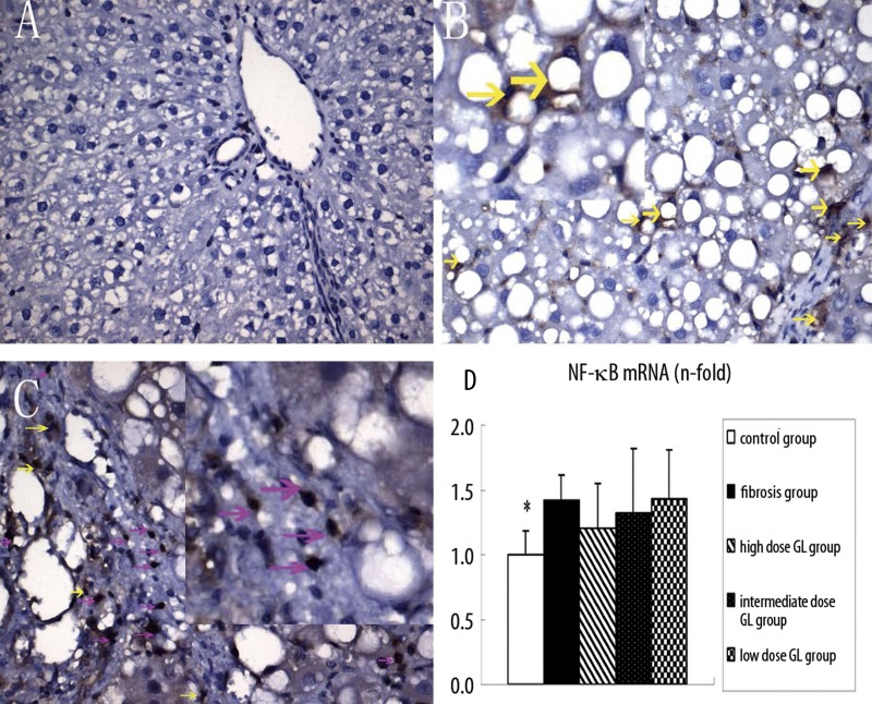
Figure 4.
Effects of 18αGL on NF-κB protein expression in rats liver tissues by immunohistochemistry staining (Positive as Brown, Original Magnification 400 (A–C), and mRNA level of NF-κB in five groups. (A–C) represented NF-κB staining of control group, liver fibrosis group, and high-dose 18αGL groups, respectively. Red arrows indicate nuclear-positive, and yellow arrows indicate the plasma positive. (D) The bargraph showed the mRNA level of NF-κB in five groups by qPCR quantity. Values are mean ± S.D; * p<0.05 vs. liver fibrosis group.

