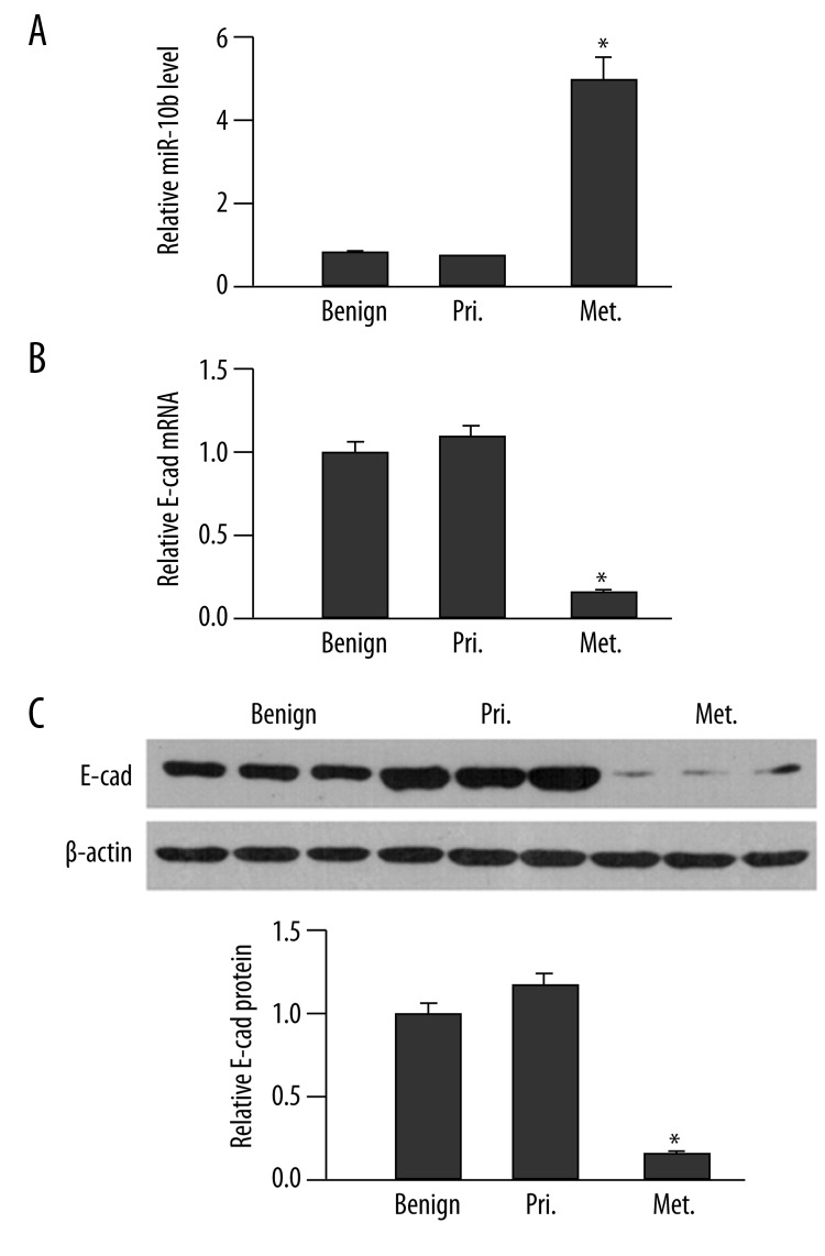
Figure 5.
miR-10b level negatively correlates with E-cad expression in human breast cancer. (A) The miR-10b level in benign breast tissues (Benign), primary breast cancers (Pri.) and metastatic breast cancers (Met.) was examined by RT-qPCR and quantified as the ratio of miR-10b to snRNA U6 (internal control), with the relative level in normal tissues arbitrarily defined as 1.0. (B) The steady-state E-cad mRNA level in indicated human tissues was determined by RT-qPCR and presented as the ratio to GAPDH (internal control), with the relative level in Mock cells arbitrarily defined as 1.0. (C) The protein level of E-cad was examined by a Western immunoblot. A representative gel image of three samples from each group is shown (top), along with quantification of the ratio of E-cad to β-actin from all tissues in each group (bottom). The level of relative E-cad expression in benign tissues was arbitrarily defined as 1.0. * P<0.05, as compared to the other two groups.

