Figures 4 and 5.


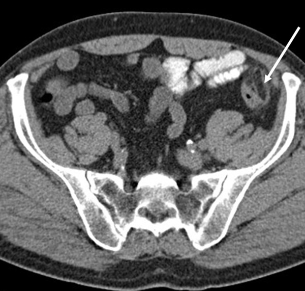
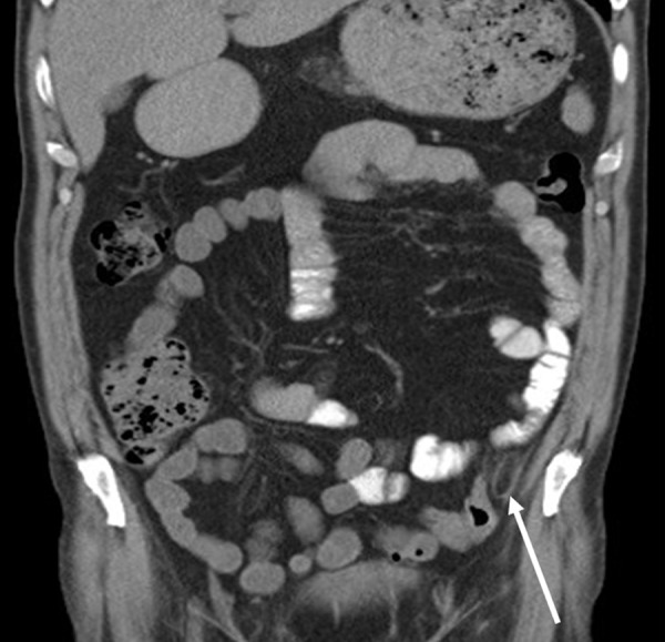
Longitudinal and transverse abdominal CT without contrast enhancement demonstrated the inflamed lesion (arrow) adjacent to the descending colon and showed an oval area with a diameter of 2.6 cm surrounded by an edematous ring.


Longitudinal and transverse abdominal CT without contrast enhancement demonstrated the inflamed lesion (arrow) adjacent to the descending colon and showed an oval area with a diameter of 2.6 cm surrounded by an edematous ring.