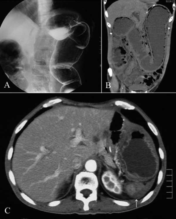
Figure 1.

(A) Contrast enema examination indicated obstruction of the large intestine in the region of the splenic flexure. (B) Coronal CT showed dilatation of the intestinal loop from the ascending colon to the transverse colon. (C) Axial CT revealed a tumor (arrow) in the dorsal region of the intra-abdominal cavity where the spleen had originally existed.
