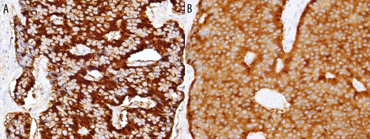
Figure 5.

Small uniform epithelial cells stained positively for chromogranin A and synaptophysin. (A) Chromogranin immunostain demonstrating the strong immunoreactivity of the tumor cells. The majority of neuroendocrine granules cluster at the base of the cells giving a prominent peripheral staining pattern. (B) Synaptophysin immunostain demonstrating the neuroendocrine nature of the tumor.
