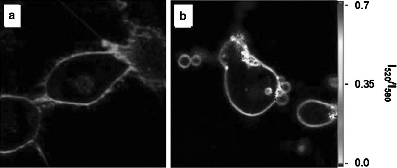
Fig. 8.
Example of an application of F2N12S in cell microscopy. Fluorescence ratiometric images of normal (a) and apoptotic (b) cells stained with F2N12S using two-photon excitation at 830 nm. Note that the images are presented in artificial color based on intensity ratio at 520 and 580 nm (Oncul et al. 2010). (Color figure online)

