
Fig. 4.
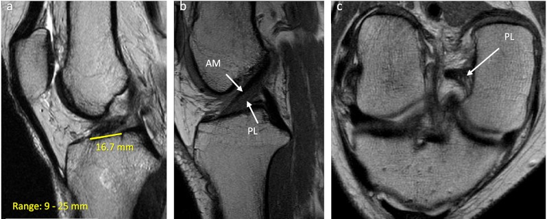
Both for diagnostic purposes and pre-surgical planning, a complete evaluation with MRI is invaluable. Regular coronal, axial and sagittal cuts should be obtained to confirm the diagnosis and look for concomitant injuries. Additionally, on the regular sagittal cuts, measurements of the insertion site length (a), inclination angle, total anterior cruciate ligament (ACL) length and quadriceps and patellar tendon thickness can be obtained. For a more precise evaluation, oblique sagittal and coronal cuts can be obtained by scanning in the same plane as the trajectory of the entire ACL. These images are particularly helpful in identifying the individual bundles (b) and potential partial bundle tears (c)