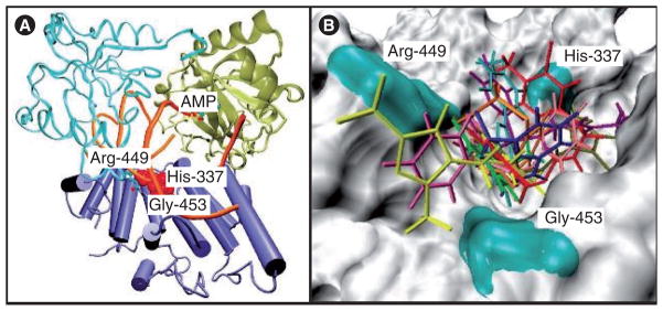
Figure 13. DNA Ligase-1.

(A) Structure of the DNA-binding domain of DNA ligase-I with the predicted binding site in red and residues His-337, Arg-449 and Gly-453 that constitute the potential binding pocket. (B) Predicted docking of some of the ligase inhibitors (Figure 14) with the DNA-binding domain [112].
