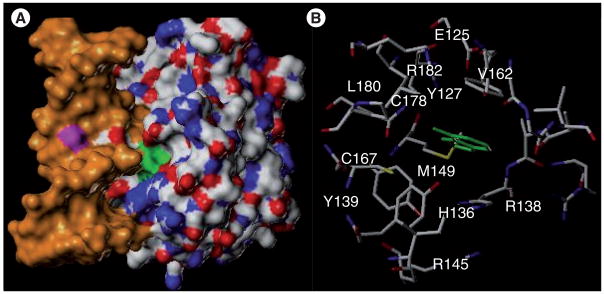
Figure 6. N-methylpurine-DNA glycosylase.

(A) Structure of human N-methylpurine-DNA glycosylase in the presence of DNA (orange) and the extrahelical hypoxanthine modification (green) buried in the enzyme’s active site. (B) View of hypoxanthine (green) substrate in active site of N-methylpurine-DNA glycosylase.
Adapted from PDB entry: 1EWN [58].
