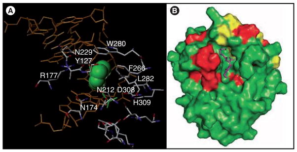
Figure 8. Human APE-1 protein.

(A) The amino acids that flank the enzymatic site of APE-1 are shown along with the DNA (orange) and the abasic site (green) adapted from PDB entry 1DEW [76]. (B) The proposed binding of E3330 (purple; see Figure 9 for structure) to APE-1 based on NMR studies (red and yellow represent amino acid residues whose chemical shifts change by >0.01 and >0.02 ppm, respectively, when E3330 is present) [84].
