Fig. 6.

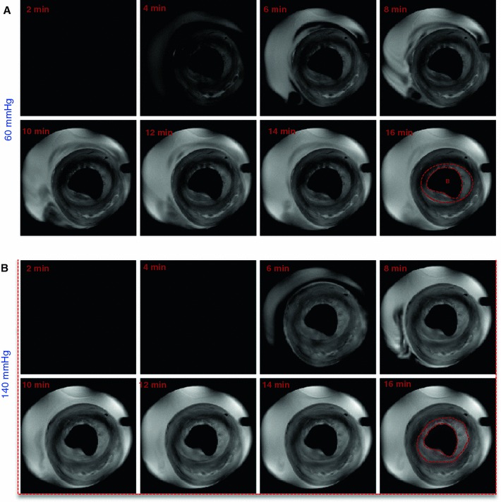
Representative T1-weighted MR images at 60 and 140 mmHg of a mid-axial slice of an isolated rat heart. Scans were acquired every 2 min after switching to Gadovist containing buffer. B balloon

Representative T1-weighted MR images at 60 and 140 mmHg of a mid-axial slice of an isolated rat heart. Scans were acquired every 2 min after switching to Gadovist containing buffer. B balloon