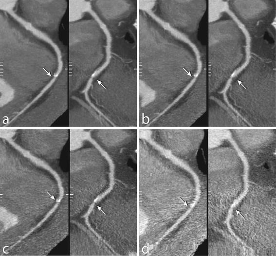
Fig. 3.

Low dose simulations of coronary CT angiography in a 64-year old male. Images show curved multiplanar reconstructions of the right coronary artery for a 100 % dose, b simulated 50 % dose, c 25 and d 12.5 % dose. A significant stenosis in the mid part of the right coronary artery (segment 3) was found (arrows) with 100 and 50 % dose. With 25 and 12.5 % dose, the stenosis was classified as “not significant”
