Fig. 2.

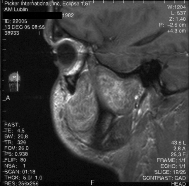
Magnetic resonance, saggital T1-weighted image after contrast administration. Large tumor in the infratemporal fossa and cheek shows intensive, inhomogenous contrast enhancement [25]

Magnetic resonance, saggital T1-weighted image after contrast administration. Large tumor in the infratemporal fossa and cheek shows intensive, inhomogenous contrast enhancement [25]