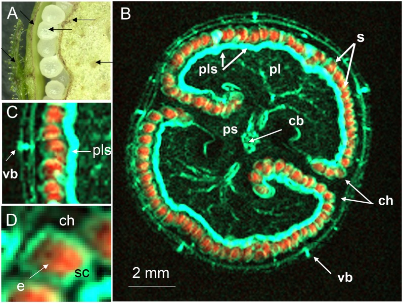
Figure 3.
Integrated MRI overlaying the water and lipid distribution within an intact tobacco capsule. Hydrated tissue colored cyan and lipid-rich tissue orange. A, Fragment of cross section (hand-dissected capsule). B, Integrated MRI image. C, Enlargement of B showing lipid-rich seeds, vascular bundle, and placental surface. D, Enlargement of B showing lipid inside of seed and water-rich testa. e, Endosperm; sc, seed coat; other abbreviations as given in the legend to Figure 2.

