Figure 4.
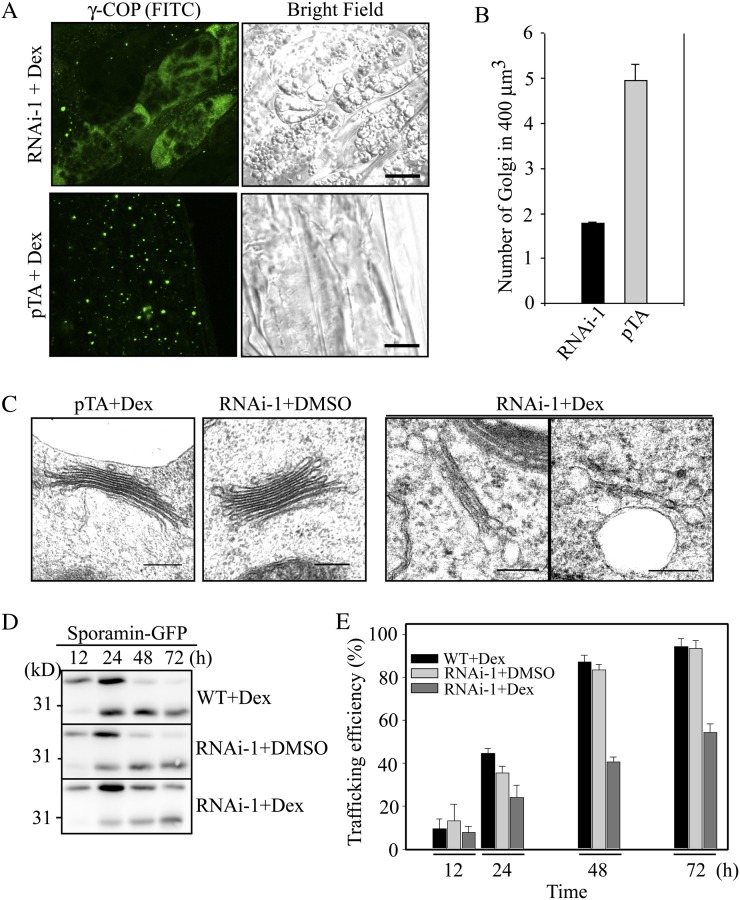
RNAi plants display severe defects in Golgi morphology and protein trafficking to the vacuole. A, Effect of RNAi on the Golgi apparatus in RNAi plants. RNAi and pTA control plants were grown on MS plates for 1 week and then transferred onto MS plates supplemented with dex (30 μm). Two days after transplanting, the plant root tissues were fixed and immunostained with γ-COP antibody (γ-COP) followed by FITC-labeled secondary anti-rabbit IgG. Bars = 10 μm. B, Quantification of the Golgi apparatus in RNAi plants. To quantify the differences in the Golgi population, serial optical z sections (10 images) obtained at 0.5-μm intervals were used for three-dimensional reconstruction using laser scanning confocal microscopy z-projection software, and the number of punctae in 400 μm3 of 50 cells was counted. Error bars indicate sd (n = 3). C, Morphological alteration of the Golgi apparatus in RNAi plants. RNAi and pTA plants treated with or without dex (30 μm) for 4 d were fixed, and ultrathin sections of root tissues were examined by electron microscopy. Bars = 200 nm. D and E, Inhibition of vacuolar trafficking of sporamin-GFP in RNAi plants. Protoplasts from RNAi that had been treated with or without dex were transformed with sporamin-GFP. D Vacuolar trafficking of sporamin-GFP was examined at various time points by western-blot analysis using a GFP antibody. As a control, protoplasts from the wild type (WT) were included. E, Trafficking efficiency was quantified using the ratio of the processed form over the total amount of expressed protein. Error bars indicate sd (n = 3).

