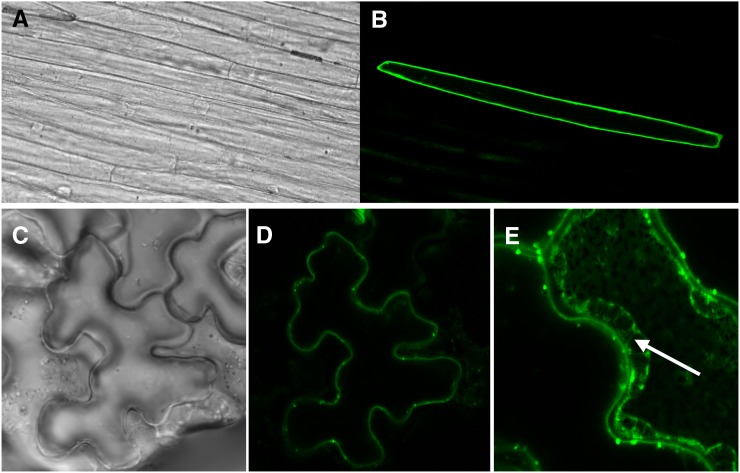
Figure 3.
TaMATE1B protein is located at the plasma membrane. GFP was fused to the N- and C-terminal ends of the TaMATE1B protein and transiently expressed in leek (A and B) and N. benthamiana (C–E) leaves. A, Bright-field image of leek tissue bombarded with a construct containing an N-terminal fusion of GFP to TaMATE1B. B, Fluorescence image of the same tissue showing GFP fluorescence around the periphery of a single cell. C, Bright-field image of N. benthamiana tissue transformed with a construct containing a C-terminal fusion of GFP to TaMATE1B. D, Fluorescence image of the same tissue showing GFP fluorescence around the periphery of a single cell. E, An N. benthamiana cell expressing the TaMATE:GFP fusion protein after plasmolysis with 100 mm Suc. The Hechtian strands that connect the cell wall with the retreating protoplast (white arrow) are consistent with TaMATE1B being located at the plasma membrane.

