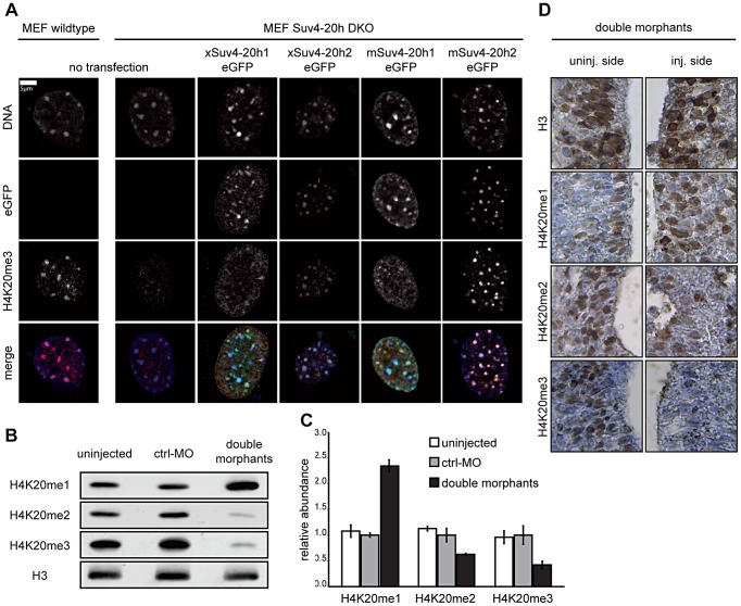
Figure 1. Functional analysis of xSuv4-20h HMTases.
(A) Transiently transfected eGFP-tagged Suv4-20h1 and h2 enzymes from frog or mouse re-establish H4K20me3 marks in heterochromatic foci of Suv4-20h1/h2 DKO MEFs. (B–D) Bulk histones from tadpoles (NF30-33) injected with morpholinos targeting translation of endogenous xSuv4-20h1 and h2 mRNA show a strong reduction in H4K20me2 and H4K20me3 levels and a concomitant increase in the H4K20me1 mark. (B) Western Blot analysis of uninjected embryos, control morphants (ctrl-MO), and double morphants with antibodies against H4K20 mono-, di- and trimethylation. PanH3 antibody was used as loading control. (C) Western Blot quantification of three independent biological experiments; data represent mean values, error bars indicate SEM. (D) Immuno-histochemistry on xSuv4-20h double morphant tadpoles. Panels show details from neural tubes stained with antibodies against the histone epitopes indicated on the side. Whole sections shown in Figure S5A.

