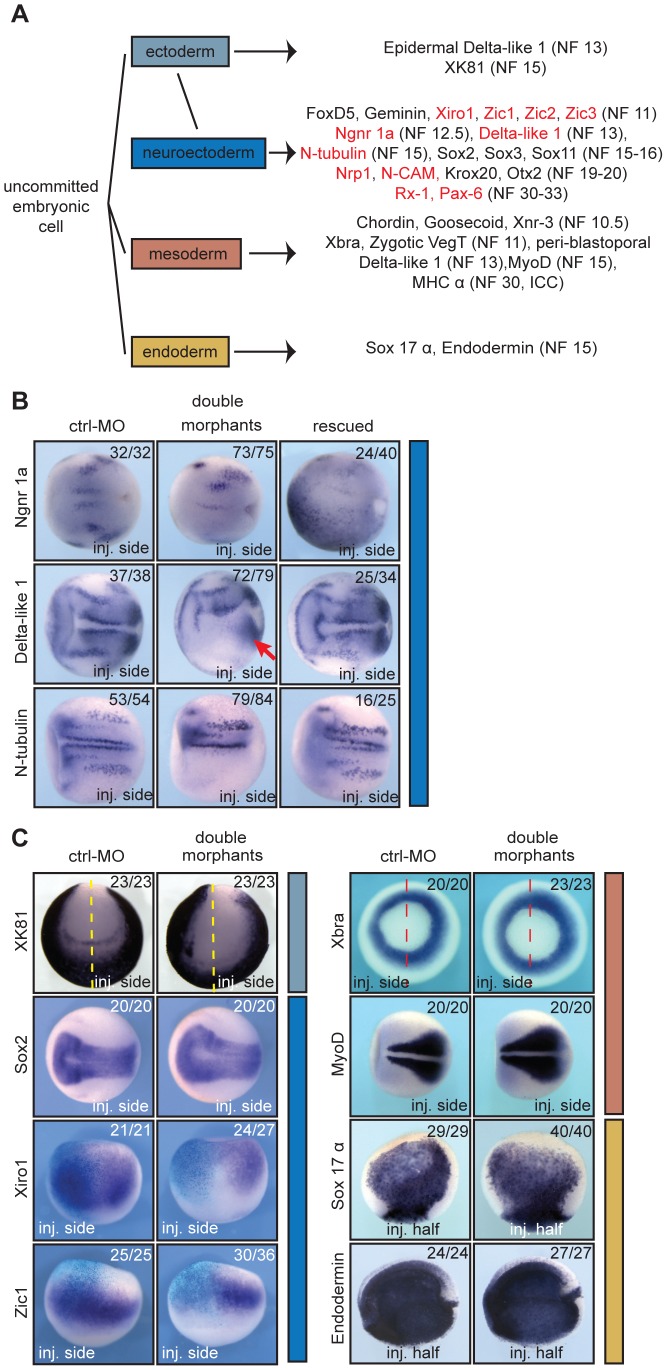
Figure 3. xSuv4-20h enzymes are required for differentiation of the neuroectoderm.
(A) Schematic illustration of analysed markers of the different germ layers (germ layer colour code extended to in situ panels). Downregulated genes upon xSuv4-20h depletion are labelled in red. (B) Expression pattern of the neuroectodermal markers Ngnr 1a (NF12.5), Delta-like 1 (NF13), and N-tubulin (NF15). The pictures show dorsal views of the open neural plate with anterior to the left. (C) Expression patterns of XK81 (ectoderm), Sox2, Xiro1, Zic1 (neuroectoderm), Xbra, MyoD (mesoderm), Sox17 α, Endodermin (endoderm) in ctrl-MO injected or double morphant embryos. XK81 - anterior views with dorsal side to the top. Sox2 and MyoD - dorsal views, anterior to the left. Xiro1 and Zic1 - dorsal views; injected halves are lineage-traced by coinjection of LacZ mRNA and subsequent β–Gal staining (light blue). Xbra - vegetal view. Sox-17 α - internal stain from the injected side in bisected embryos, animal pole up. Endodermin – internal stain from the injected side in bisected embryos; anterior to the left.

