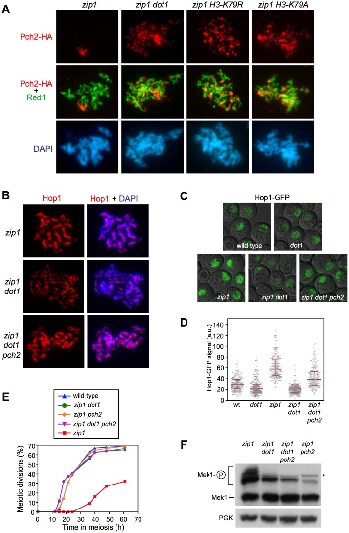
Figure 7. H3K79me controls Hop1 localization by excluding Pch2 from chromosomes.
(A) H3K79me is required to prevent Pch2 localization outside of the rDNA. Immunofluorescence of meiotic chromosome spreads stained with DAPI (blue), anti-HA (red) and anti-Red1 (green) antibodies. Strains are: DP1050 (zip1), DP1053 (zip1 dot1), DP1052 (zip1 H3-K79R) and DP1051 (zip1 H3-K79A). (B–D) The absence of Pch2 partially restores Hop1 chromosomal abundance in zip1 dot1. (B) Immunofluorescence of meiotic chromosome spreads stained with DAPI (blue) and anti-Hop1 antibody (red). Strains are: DP428 (zip1), DP655 (zip1 dot1) and DP1054 (zip1 dot1 pch2). (C) Representative images of cells expressing HOP1-GFP in wild type (DP963), dot1 (DP966), zip1 (DP964), zip1 dot1 (DP965) and zip1 dot1 pch2 (DP1027). (D) Quantification of the Hop1-GFP signal intensity on fluorescence images (a.u., arbitrary units). 300 individual nuclei were analyzed for each strain. Each spot in the plot represents the fluorescence intensity of every nucleus measured. Error bars represent the median with interquartile range. P<0.01 in pairwise comparisons. In all cases (A–C), spreads were prepared and GFP images were taken 24 h after meiotic induction in ndt80 strains. (E, F) The absence of Pch2 does not restore the pachytene checkpoint response in zip1 dot1. (E) Time course of meiotic nuclear divisions; the percentage of cells containing more than two nuclei is represented. Strains are: DP421 (wild type), DP422 (zip1), DP555 (zip1 dot1), DP1029 (zip1 pch2) and DP1041 (zip1 dot1 pch2). (F) Western blot analysis of zip1-induced Mek1 phosphorylation in ndt80 strains. PGK was used as a loading control. The asterisk marks a presumed non-specific band (see Figure 3D). Strains are: DP428 (zip1), DP655 (zip1 dot1), DP881 (zip1 pch2) and DP1054 (zip1 dot1 pch2).

