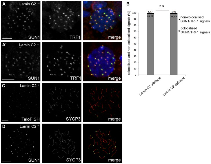
Figure 3. Loss of lamin C2 has no effect on meiotic telomere attachment.
(A) 3D-preserved swab preparations showing wildtype (A) and knockout (A′) spermatocytes simultaneously labelled with anti-TRF1 and SUN1 antibodies. As in the wildtype, in lamin C2−/− spermatocytes virtual all telomeres appear to be attached to the NE as indicated by co-localisation of TRF1 and SUN1 signals. Scale bars 5 µm. (B) Quantifications of co-localised and non-co-localised TRF1/SUN1 signals (see A) revealed that ratios of co-localised to non-co-localised spots comparing wildtype and knockout spermatocytes show no significant difference (wildtype n = 33; lamin C2−/− n = 45; Pearson's Chi2 test p-value: 0.799). (C,D) Chromosome spread preparations of pachytene-like lamin C2−/− spermatocytes showing that all telomeres are associated with SUN1. In (C) TeloFISH and in (D) anti-SUN1 staining in co-localisation with SYCP3. Scale bars 10 µm.

