Figure 4.
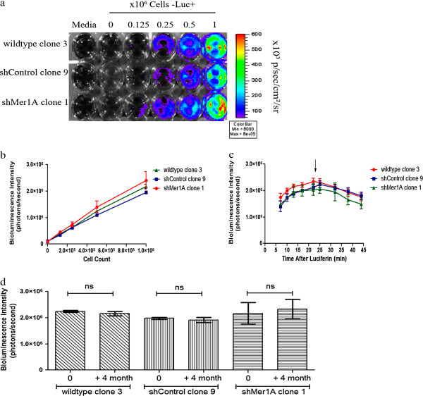
In vitro quantitation of bioluminescence signal in monoclonal human leukemia cell lines expressing luciferase.a Pseudocolor representation of the bioluminescence intensity from monoclonal luciferase-transduced Jurkat cell lines (wildtype, shControl, shMer1A). Cell concentrations ranging from 1.25 x 10⁵ to 1 x 10⁶ cells were plated in a 24-well plate and images were captured after addition of D-luciferin to the media. Wells containing medium only with or without D-luciferin served as negative controls. b Correlation between cell number per well and bioluminescence intensity (photons/second per well) for three cell line derivatives. Mean values (+/− SEM) were determined from three separate experiments. The measured intensity of bioluminescence was directly proportional to the number of cells. c Bioluminescence intensity as a function of time after luciferase addition in monoclonal luciferase-transduced Jurkat cell lines (wildtype, shControl, shMer1A). Mean values and standard errors (+/− SEM) were derived from three independent experiments. No significant differences in the dynamics of signal intensity over time were observed for the selected clones. d Stability of luciferase activity of three monoclonal populations of the Jurkat cell line (wildtype, shControl, shMer1A). Cells were passaged for four months and luciferase activity was monitored by measurement of bioluminescence intensity. Mean values (+/− SEM) derived from three independent measurements. All clones exhibited stable luciferase activity throughout the test period.

