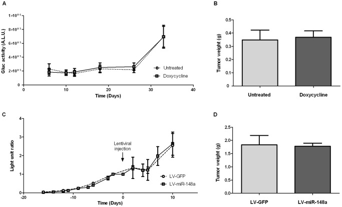
Figure 4. Murine orthotopic xenograft and tumor progression monitoring.
(A) In vivo monitoring of xenograft tumor progression: MIA PaCa-2 cells expressing miR-148a and secreted Gaussia luciferase (Gluc) were injected in the pancreas of SCID mice. Mice received normal water (untreated, n = 10) or water supplemented with doxycycline (doxycycline, n = 12) for miR-148a expression induction until sacrifice. Gluc levels were measured in mice serum. The results are the mean of Gluc activities (±SEM) and are expressed as Arbitrary Light Unit (A.L.U.). (B) Tumors were removed and weighted the day of surgery. Results are mean (±SEM) of tumor weight in the untreated group (n = 10) and the doxycycline treated group (n = 12) (C) In vivo monitoring of xenograft tumor progression: MIA PaCa-2 cells expressing secreted Gaussia luciferase were injected in the pancreas of SCID mice (n = 10). Fifteen days later, lentiviral vectors encoding miR-148a (miR-148a, n = 7) or GFP only (GFP, n = 3) were injected in the tumors (the arrow indicates the lentiviral particles injection). The amount of Gluc was measured in mice serum and compared to the Gluc amount measured the day of lentivector injection (Day 0). The results are the mean of the Gluc level ratio in each group (±SEM) and are expressed as arbitrary light unit ratio. (D) Tumors receiving miR-148 (miR-148a, n = 7) or GFP (GFP, n = 3) were removed 10 days after lentiviral injection and were weighted. The graph represents the comparison of tumor weight expressing miR-148a (n = 7) or GFP (n = 3). The results are the mean of tumor weight in each group (±SEM).

