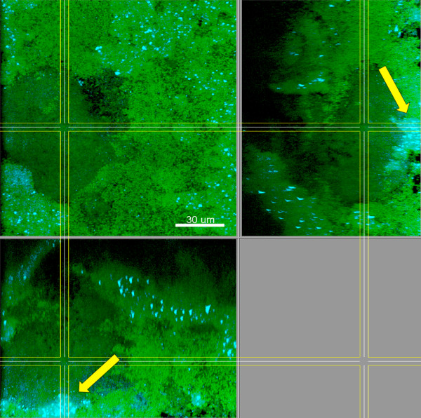
Figure 6.
Biofilms grown for 64.5 h in iHS medium. FISH staining of a fixed biofilm; the biofilm base in the side views is directed towards the top view. Cyan: V. dispar, green: non-hybridised cells, DNA staining (YoPro-1 + Sytox). Arrows: Microcolonies of V. dispar. Shown is a representative area of one disc. Scale bar: 30 μm.

