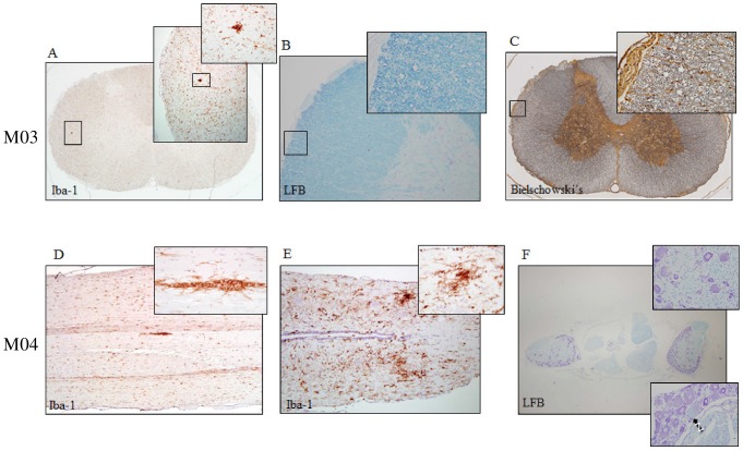
Figure 7. Spinal cord pathology in two HHV-6A intravenously inoculated marmosets.
Iba-1 is specific for microglia and macrophages, Luxol Fast Blue (LFB) stains myelin and Bielschowski's stains neurofibrils. Cervical spinal cord pathology of M03 includes (A) microglial/macrophageal aggregates identified by Iba-1, and swollen myelin sheaths identified by (B) LFB and (C) Bielschowski's. Spinal cord pathology of M04 includes microglial/macrophageal aggregates identified by Iba-1 in the (D) thoracic and (E) lumbar spinal cord and (F) myelin abnormalities identified by LFB in the dorsal root ganglia, specifically variations in sheath size and focal neuronal chromatolysis (black arrow), indicative of mild reversible damage.

