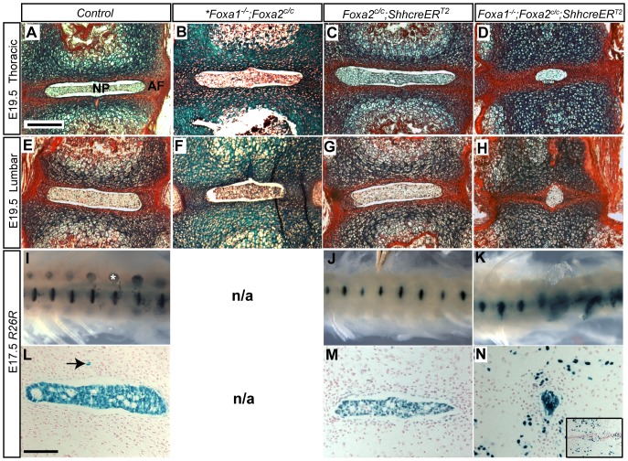
Figure 2. Foxa1;Foxa2 double mutants have severe defects in intervertebral disc formation.
A–H: Alcian blue and picrosirius red staining of disc sections. Scale bar is 200 µm. Thoracic sections at E19.5 show a normal sized nucleus pulposus (NP) surrounded by a red-stained annulus fibrosus (AF) in control (A) Foxa1 null (B) and Foxa2 notochord knockout (C) embryos. Asterisk in Foxa1−/−;Foxa2c/c column denotes that embryo was E17.5. Disc morphology was also normal at the lumbar region in these mice (E, F, G). In Foxa1;Foxa2 double mutants (D and H) nuclei pulposi were abnormal. Nuclei pulposi were observed to be much smaller than nuclei pulposi present in control or individual single mutants. I and J: A fatemap of notochord cells using the R26R reporter allele demonstrated proper nuclei pulposi formation in control (I) and Foxa2 notochord knockouts (J). In contrast, Foxa1;Foxa2 double mutants (K) had severely deformed nuclei pulposi and blue cells were dispersed. Defects were more severe in the posterior. (I) is an embryo of the genotype Shhgfpcre;R26R. White asterisk marks dorsal root ganglia. The Shhgfpcre allele is constitutively active, therefore all Shh-expressing cells, including dorsal root ganglia, and their descendants are LacZ-positive. All LacZ positive cells in J and K are derived exclusively from the notochord [3]. In our mating it was not possible to obtain a Foxa1−/− notochord fatemap without also removing the floxed Foxa2 allele, therefore the Foxa1−/− fatemap is not shown (N/A = not applicable). L, M and N: Sections of LacZ-stained tissue at the lumbar level. Inset in N shows an additional section outside of the plane of the NP; numerous cells are scattered throughout the disc region and vertebral bodies. L–M: Scale bar is 100 µM. Arrow in L denotes a notochord remnant, which is a notochord cells that does not reside in the NP.

