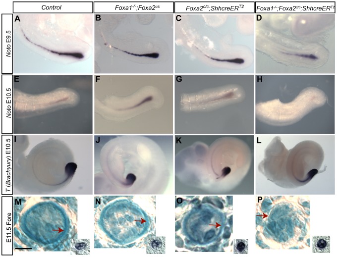
Figure 5. Expression of some notochord genes are unaffected in Foxa1;Foxa2 double knockouts but the notochord sheath is abnormal.
A–D: E9.5 and E–H: E10.5 whole-mount in situ hybridization for Noto. I–L: E10.5 whole-mount in situ hybridization for T (Brachyury). Both genes are expressed in the posterior notochord and expression extends through the tail of the embryo. Noto expression at E9.5 in single (B,C) and double mutants (D) was indistinguishable from control embryos (A). However, at E10.5, Noto was barely detectable in the tail of double mutants (H). I–L: T (Brachyury) expression at E10.5. There was no detectable differences in T expression in control (I), single mutant (J,K) and double mutant embryos (L). E11.5 alcian blue and nuclear fast red staining of the notochordal sheath at the forelimb level (M–P). Scale bars are 10 µm. Adjacent or near-adjacent sections of the notochord are pictured in the inset of M–P and show T (Brachyury) in situ hybridization of the notochord. At the forelimb level, the notochord sheath is clearly visible as a thin blue ring around the notochord in control (M) and single mutant (N,O) embryos (red arrows). In double mutants, the notochord is visible but the sheath is barely apparent (P, red arrow).

