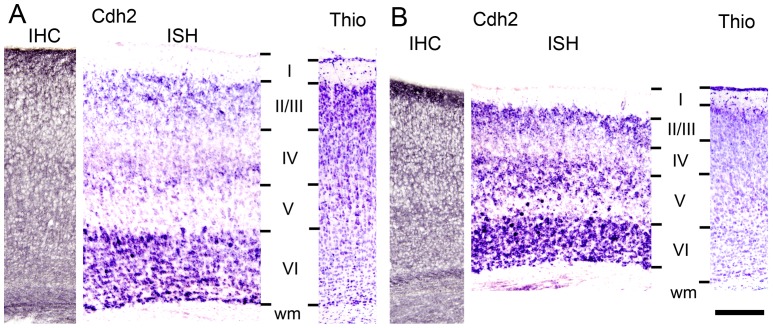
Figure 5. In vivo expression of N-cadherin in the cerebral cortex during postnatal mouse development.
(A, B) Immunohistochemistry (IHC) and in situ hybridization (ISH) to detect expression of N-cadherin (Cdh2) at the protein level and mRNA level, respectively. Coronal sections of the somatosensory cortex (A) and the visual cortex (B) of the mouse at postnatal day 6. Note the almost complete lack of N-cadherin mRNA expression in layer V somatosensory neurons (A, ISH) and the relatively strong expression in a subpopulation of layer V visual neurons (B, ISH). IHC did not show layer-specific expression, because pyramidal cell dendrites extend over several layers. For identification of cortical layers, a corresponding Nissl stain (Thio) is shown in the right panels. Other abbreviations: I–VI, cortical layers I–VI; wm, white matter. Scale bar: 200 µm (for A, B).

