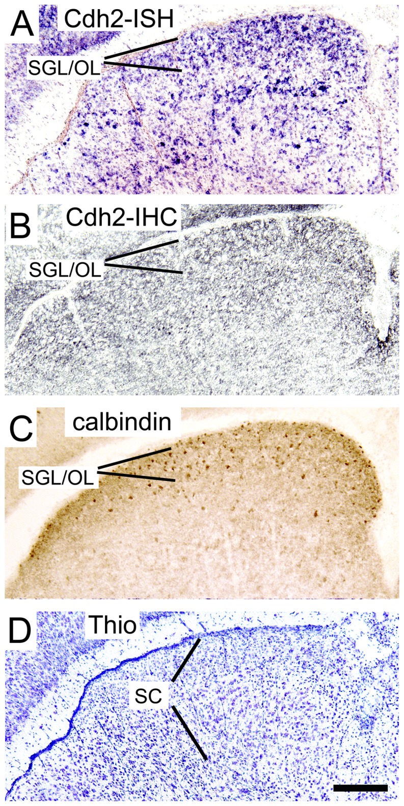
Figure 6. In vivo expression of N-cadherin in the superior colliculus.

(SC) during postnatal mouse development. (A) In situ hybridization (ISH) to detect N-cadherin (Cdh2) mRNA expression. Note the strong expression of N-cadherin in neurons of the superficial collicular layers (prospective superficial gray layer [SGL] and optic layer [OL]) where most visual cortical axons terminate. (B, C) Immunohistochemistry (IHC) to detect N-cadherin protein (B) and calbindin D28k protein (C), a marker for SGL and OL. (D) Nissl stain of a section adjacent to that shown in A and B. Coronal sections through the postnatal day 6 superior colliculus are shown. Scale bar: 200 µm (for A–D).
