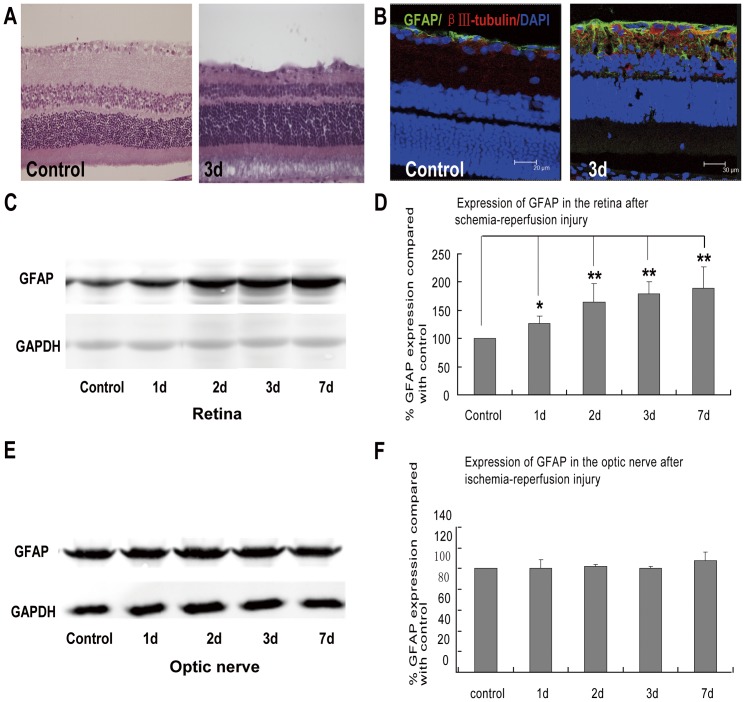
Figure 1. Different GFAP expression between retina and optic nerve after the induction of transient intraocular hypertension.
(A) HE staining showed that comparing with the intact and regular retina in control rats, transient intraocular hypertension induced significant decrease in the thickness of retina as early as 3 days. (B) In control retina, GFAP immunoreactivity was confined to the nerve fiber layer (NFL) and ganglion cell layers. Three days after the induction of transient intraocular hypertension, GFAP-labeled processes were found to enwrap the RGCs and extend throughout the entire retina. (C–D) Immunoblots revealed increased expression of GFAP in the retina at 1, 2, 3, and 7 days after the injury. (E–F) While in the optic nerve, the expression of GFAP seemed to be constant throughout the observation course. * p<0.05; ** p<0.01.

