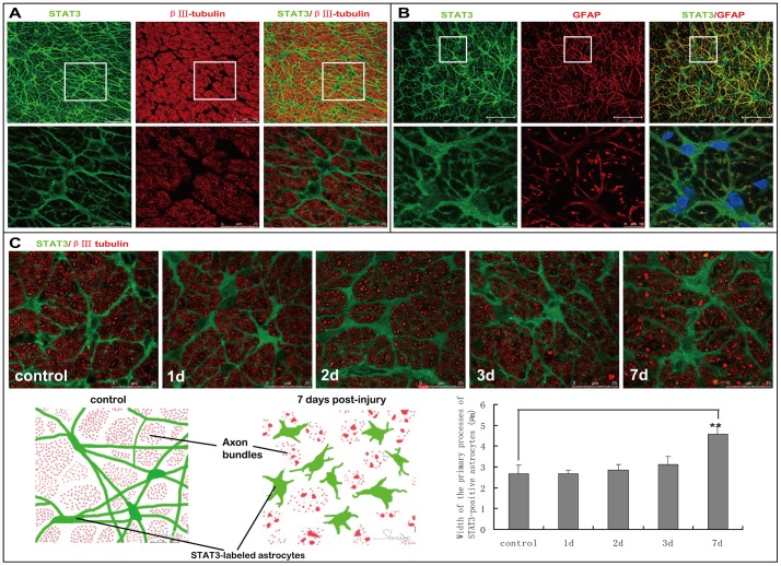
Figure 3. Expression of STAT3 in the astrocytes of normal and transient intraocular hypertension-injured optic nerve.
(A) In control rats, STAT3 immunoreactivity was observed in the processes and perinuclear cytoplasm of astrocyes. STAT3-positive processes connected to form an appearance of honeycomb and partitioned neighboring RGC axons into bundles. Spaces among these axon bundles were perfectly filled by STAT-3 positive cell body. (B) STAT3 showed good co-localization with GFAP in the processes of astrocytes. However, STAT3-positive signal could also be observed at the perinulear cytoplasm, where no GFAP immunoreactivity was found. Therefore STAT3 may give clearer and more entire appearance of the astrocytes than GFAP. (C) From day 3 after the induction of transient intraocular hypertension, STAT3-labeled primary processes of astrocytes in the optic nerve became thicker and tortuous,destroying normal regular honeycomb architecture of the glias. At day 7, hypertrophy of cell soma, thickening of primary processes, and retraction of higher-order processes in STAT3-positive astrocytes were easily identified. The schematic in C depicted how the morphology of STAT3-positive astrocytes changed following transient intraocular hypertension. Quantitative analysis showed that average thickness of STAT3-labeled primary processes was 4.58±0.36 µm at day 7 post-injury compared with 2.68±0.41 µm in normal optic nerve.

