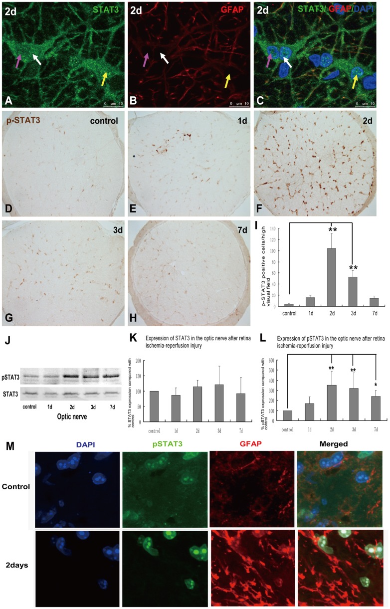
Figure 4. Expression of pSTAT3 in control and transient intraocular hypertension injured optic nerve.
Nuclear translocation of STAT3 was observed in the optic nerve at 2 days after the induction of transient intraocular hypertension, which may suggest the activation of STAT3 signalling pathway in this injury (See the arrows in A–C). One day after the induction of transient intraocular hypertension, a small amount of pSTAT3-positive cell nuclei was observed in the central region of the optic nerve. Significant increase of p-STAT3-immunolabeled signal was detected throughout the optic nerve at 2 days and was markedly decreased at 3 and 7 days (D–H). Counting of p-STAT3 -positive nuclei was shown in I. Western blot revealed that both STAT3 and pSTAT3 were mildly expressed in the optic nerve of control rats. PSTAT3 expression was substantially increased at 2 days after transient intraocular hypertension and this increase was markedly attenuated but still higher than control optic nerve at 7 days. Expression of STAT3 kept relative consistent before and after the induction of injury (J). See the quantitative analysis in K and L. Double immunofluorescence labeling for pSTAT3 and GFAP in optic nerve sections in control and Day 2 post-ischemic reperfusion injury. (Blue: DAPI; Green, anti-phosphorylated STAT3; Red, anti-GFAP) (M).

