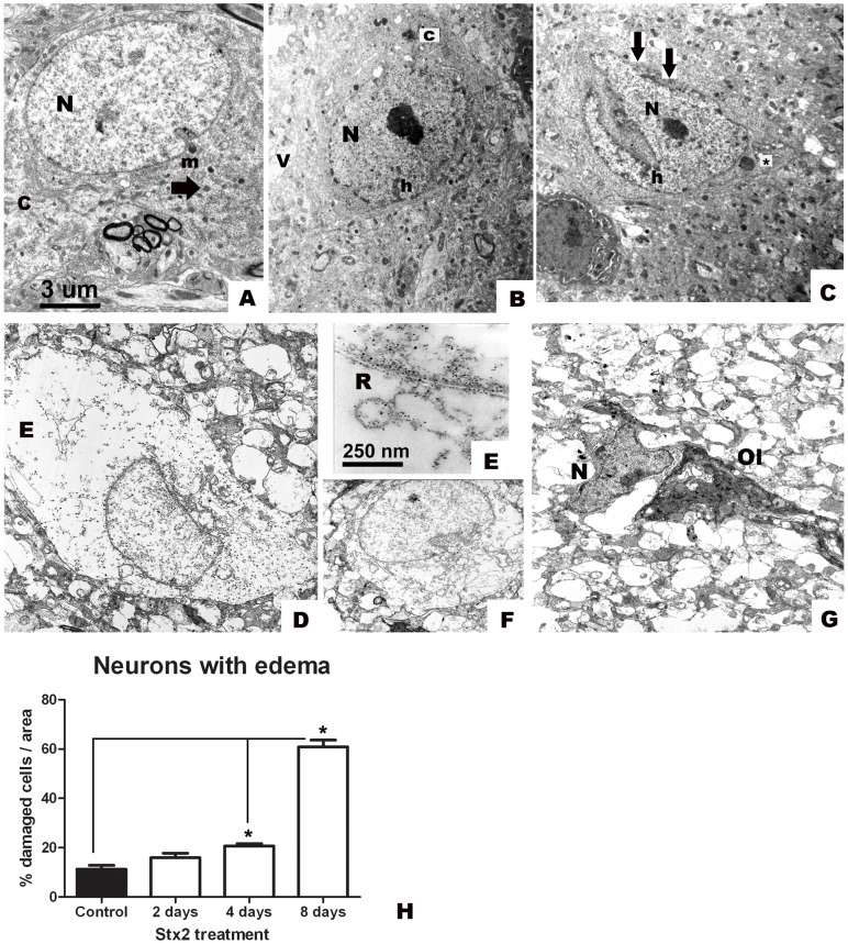
Figure 2. Intravenous administration of Stx2 causes neuronal damage.
Conserved striatal neuron after i.v. vehicle (saline) administration (A); pale nucleus (N) and intact cytoplasm (c) and membranes (m); intact mitochondrion (arrow). After 2 days of treatment: vacuolated neuron with contrasted cytoplasm (c) and nucleus (N) (B); heterochromatin condensation (h). After 4 days: more contrasted nucleus (N) with increased heterochromatin (h) condensation and prominent indentation with loss of membranes (arrows) (C); vacuolated perinuclear mitochondrion (*) (C). Neuron with edema and loss of regular nuclear shape on day 8 (D). Disorganized endoplasmic reticulum (R) in the cytoplasm with edema (E) at higher magnification; loss of regular shape in the nucleus (F) of a neuron with edema (E); affected neuronal nucleus (N) with irregular shape and no apparent surrounded cytoplasm; (Ol, oligodendrocyte) (G). These features are absent in striatal neurons of the vehicle-treated group (A). Quantification of the percentage of damaged neurons with edema (H). Significant results are observed starting on day 4. Maximum number of neurons with edema in Stx2-treated mice observed on day 8 (*) (H). Results are expressed as a percentage of the total number of neurons in an area of 3721 µm2. Data are mean ±SEM of 6–8 experiments (F and G). An asterisk denotes statistical significance, p<0.05. The scale bar in A applies to micrographs B–D and F–G.

