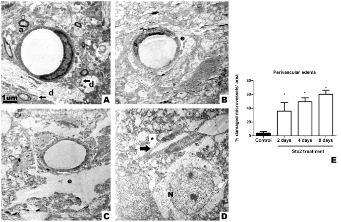
Figure 4. Intravenous administration of Stx2 causes ultrastructural alterations at the blood brain barrier level.
Electron micrograph showing a conserved endothelial cell with conserved endothelial nucleus that constitutes a microvessel from a striatal brain slice after i.v. administration of vehicle; microvessel surrounded by conserved synapses (arrows), dendrites (d) and myelinated axons (a) (A). Perivascular edema (e) 2 days after i.v. administration of Stx2 (B). After 4 days: more pronounced perivascular edema (e) (C). After 8 days: infarcted microvessel (arrow) with perivascular edema (*) near a neuron (N) (D). No cytoplasmic membrane is observed in the neuron at this stage. Quantification of the percentage of damaged microvessels by perivascular edema (E). Damage in microvessels begins to be significant on day 2 and is maximum on day 8 (*). Results are expressed as a percentage of the total number of microvessels in an area of 3721 µm2. Data are mean ±SEM of 6–8 experiments (G). An asterisk denotes statistical significance, p<0.05. The scale bar in A applies to micrographs B–D.

