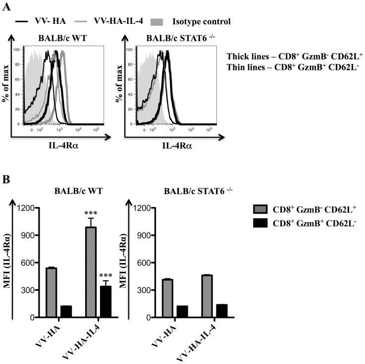
Figure 6. IL-4Rα is up-regulated on CD8+ T cells in a STAT6 dependent manner following VV-HA-IL-4 infection.
BALB/c WT mice (n = 5) or BALB/c STAT6 −/− mice (n = 5) were infected with 5×10⁶ PFU of VV-HA control, VV-HA-IL-4 or kept unimmunized for 7 days prior to sacrifice and analysis using flow cytometry. A and B, Representative histogram plots showing cell surface IL-4Rα expression (A) and the mean (n = 5) MFI of cell surface IL-4Rα expression (B) on the indicated CD8+ splenocyte subset from infected mice of the indicated genetic background. The data shown is representative of two independent experiments and the error bars depict the SEM. One-way ANOVA (Tukey's Multiple Comparison) was used to determine the statistical significance of the data relative to respective cell subset from VV-HA infected mice (*** - p<0.001).

