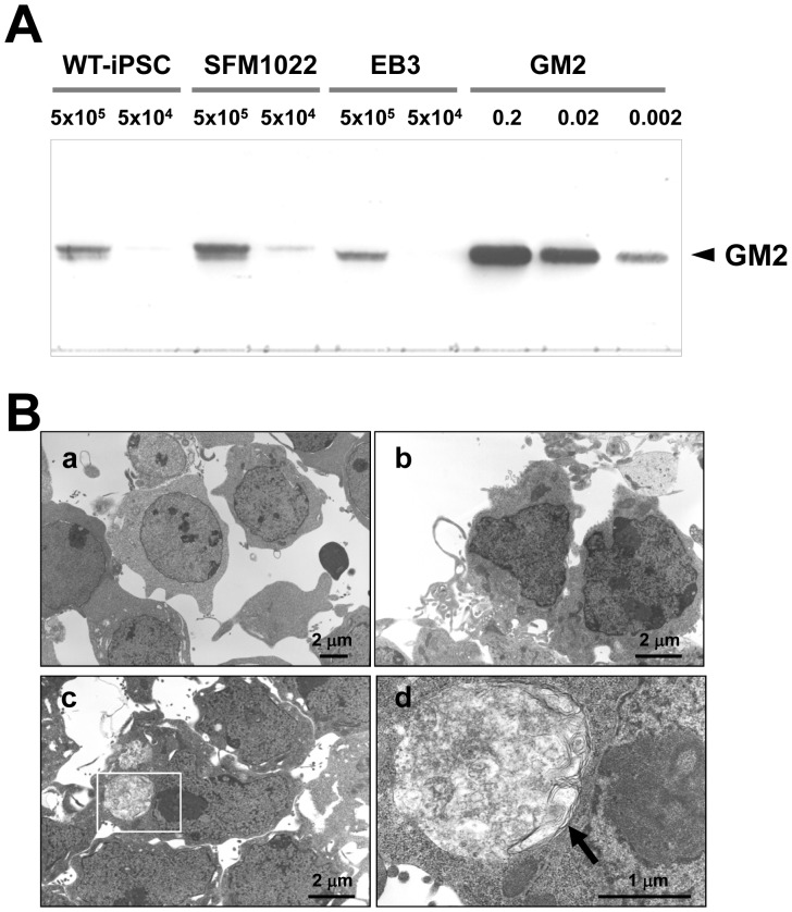
Figure 5. Disease phenotypes of iPSCs.
A, TLC immunostaining, with anti-GM2 antibodies, of SFM1022, EB3 cells and WT-iPSCs. Authentic GM2 ganglioside (0.002, 0.02 and 0.2 µg) was used as a control. B, Electron micrographs of differentiated neural cells derived from EB3 cells ( a), WT-iPSCs (b), and SFM1022 cells (c, d). The boxed region in c is magnified in d. Inclusion bodies with a membranous and pleomorphic shape (arrow) were identified in the cytoplasm of SFM1022 cells, but not in that of EB3 cells and WT-iPSCs (a: ×3000, b: ×5000, c: ×5000, d: ×20000).

