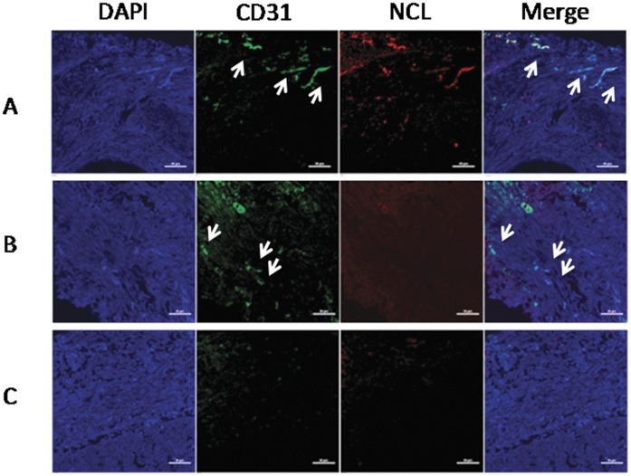
Figure 1. The NSCLC sections were divided into three groups based on the blood vessel density and nucleolin expression.
The tumor blood vessels and nucleolin were stained with anti-CD31 (green) and anti-nucleolin (red), respectively. Nuclei were stained with DAPI (blue). Representative images from the three groups were shown here. (A) CD31hiNCLhi, (B) CD31hiNCLlo, (C) CD31loNCLlo. Scale bar, 50 µm.

