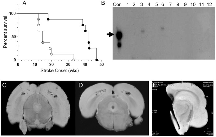
Figure 4. Juvenile EPC-therapy delays stroke onset.
A) Survival curve analysis comparing mock-treated controls (○) and EPC-treated (•) spTg25+ female rats, P = 0.003. B) PCR-analysis for Y-chromosome DNA detected the expected Y-chromosome sequence-specific 104 bp product in the positive control lane (Con) and in EPC-treated rat brain microvessels at 1 week (lane-3) and 2 weeks (lane-6) after EPC infusion. No Y-DNA was detected at 3- (lane 9) and 4-weeks (lane 12) after EPC-infusion. Y-DNA was not detected in other tissues tested: kidney (lanes 1,4,7,10), and whole brain (lanes 2,5,8,11). C-E) Representative images of gradient-echo MRI analyses of rat brains from: C) a mock-treated rat at stroke onset, D) an EPC-treated rat at significantly-delayed stroke onset, and E) a half-brain from an asymptomatic, EPC-treated rat 3-weeks after EPC-infusion.

