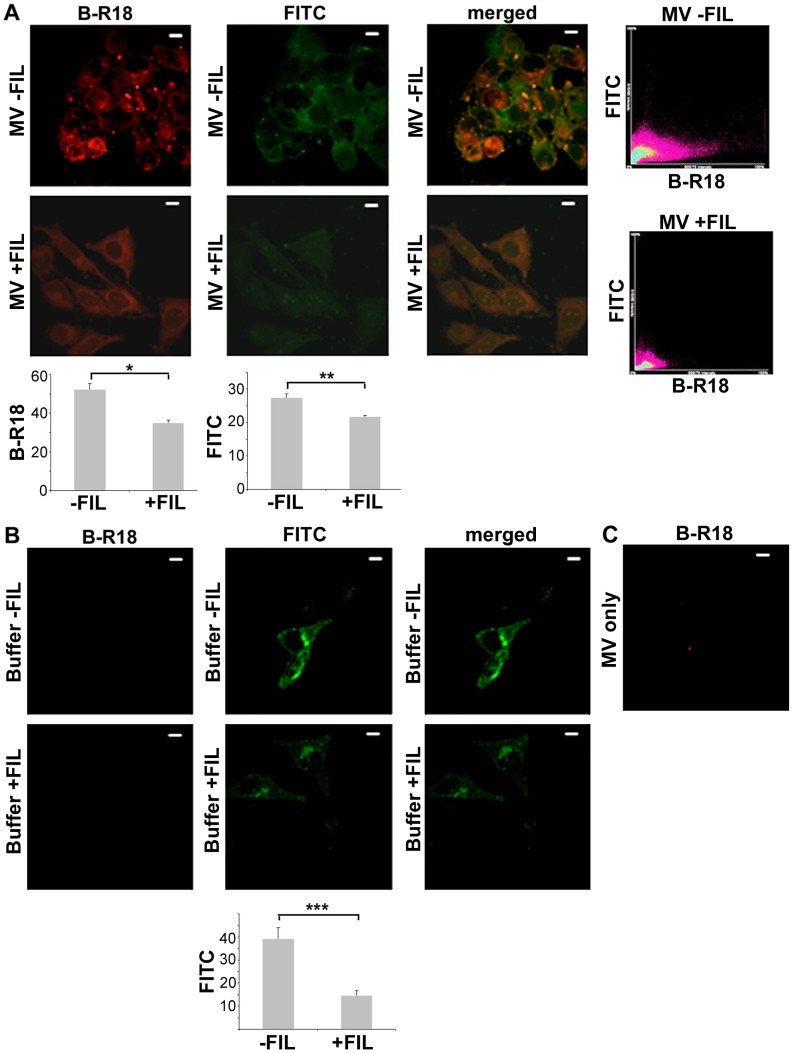
Figure 7. Cholesterol-dependent fusion of S. aureus MVs with HeLa cells.
Localization of rhodamine B-R18-labeled MVs (red fluorescence used as a readout for MV fusion with the host cell plasma membrane), and FITC-conjugated lipid raft marker CtxB (green fluorescence) in HeLa cells after 30 min of incubation with membrane-derived vesicles obtained from strain 8325-4 (MV; panel A), and with PBS (buffer; panel B). Treatment was done in the absence (−FIL) and in the presence (+FIL), respectively, of the cholesterol-sequestering agent Filipin III (final concentration 10 µg/ml). Bar graphs show quantitative analysis of red (B-R18) and green (FITC) fluorescence in treated HeLa cell samples. Values represent arbitrary units of pixel intensity for red and green fluorescence determined using ImageJ, and shown are the means ± SEM of data collected from 10 cells. *P = 0.0001, **P<0.0001, and ***P = 0.0002, for treatment in the absence vs presence of Filipin III. The merged images show the labeling with both fluorescent dyes. The scattergrams in panel A with red (B-R18) and green (FITC) pixels plotted on graphs were used to obtain the colocalization coefficient (rp) between MVs and CtxB in the HeLa cells treated with strain 8325-4 MVs for 30 min. (C) B-R18-labeled MVs alone. Magnification: 1000×. Bars = 10 µm.

